
Neurosurgery remains one of the most demanding disciplines in modern medicine, where a single millimeter of deviation can mean the difference between a successful recovery and permanent neurological deficit. Surgeons traditionally rely on pre-operative scans to map out a path through the delicate folds of the brain, yet the brain shifts significantly once the skull is opened, rendering those initial maps less accurate as the procedure progresses. To address this intraoperative variability, the medical community has turned toward the integration of robotic assistance directly within the magnetic resonance imaging environment. This convergence aims to provide a continuous loop of high-resolution data and robotic precision that adjusts to real-time physiological changes. By removing the guesswork associated with brain shift and providing a stable, tremor-free interface for tool manipulation, these systems represent a fundamental shift in how complex intracranial pathologies are approached today.
Integrating Robotic Systems within Magnetic Resonance Environments
Technical Hurdles: Overcoming Magnetic Interference
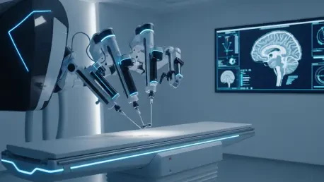
The primary challenge in combining these two powerful technologies lies in the intense electromagnetic environment of a standard MRI suite. Traditional surgical robots are constructed from ferromagnetic materials and rely on electric motors that generate electromagnetic interference, which would typically distort MRI images or even turn the robot into a projectile. Engineers have overcome these obstacles by utilizing non-ferrous materials such as high-grade ceramics, carbon fiber, and specialized polymers that do not react to the strong magnetic fields. Instead of standard electric motors, these advanced systems often employ piezoelectric actuators or pneumatic power sources that function flawlessly without introducing artifacts into the imaging data. This meticulous selection of materials ensures that the surgical arm can operate within the bore of the magnet while the scanner continues to produce crystal-clear images of the operative site.
Mechanical Synergy: Integrating Logic and Motion
Beyond the mechanical components, the software integration required to sync robotic movements with the high-speed pulse sequences of the MRI is equally complex. Digital control systems must be shielded against radiofrequency noise to prevent signal degradation that could obscure the surgical field. Recent developments in 2026 have introduced low-latency data processing units that can interpret the scanner’s output and update the robot’s position within milliseconds. This rapid synchronization is essential when navigating near the brainstem or major vascular structures, where even minor pulsations from the patient’s heartbeat must be accounted for by the robotic compensators. By establishing a robust communication link between the imaging hardware and the surgical actuator, these systems provide a level of oversight that manual surgery cannot match. This technological synergy allows for a more confident approach to pathologies previously deemed too risky.
Transforming Patient Care and Surgical Paradigms
Precision Engineering: Enhancing Accuracy through Feedback Loops
Operating with real-time MRI guidance allows for a closed-loop system where the robotic arm adjusts its trajectory based on the most recent anatomical data. In conventional setups, a surgeon might use a static frame-based system, but MRI-integrated robotics allow for dynamic tracking of deep-seated targets such as tumors or specific nuclei for deep brain stimulation. As the robot advances a probe or a laser ablation catheter, the scanner provides thermal maps or diffusion-weighted images to show exactly how the tissue is responding in the moment. This capability is particularly vital for treating glioblastomas, where the borders of the tumor are often indistinguishable from healthy brain matter to the naked eye. The robotic system maintains a steady hand, following the digital boundaries defined by the surgeon on the live scan, which significantly reduces the risk of accidental injury to critical functional zones.
Future Directions: Broadening Applications and Global Access
Looking ahead from 2026 to 2030, the scope of MRI-compatible robotics is expected to expand into pediatric neurosurgery and the treatment of complex vascular malformations. The precision offered by these systems is uniquely suited for the smaller, more delicate structures found in pediatric patients, where the margin for error is even slimmer than in adults. Additionally, the integration of artificial intelligence with these robotic platforms is beginning to allow for predictive modeling of brain shift before it occurs, further refining the accuracy of the guidance systems. Institutions adopting these technologies are finding that the initial high cost is offset by the reduction in long-term complications and the ability to treat previously inoperable cases. As the hardware becomes more compact and user-friendly, smaller regional centers may soon be able to offer the same level of care once reserved for major academic research hospitals.
The integration of robotic precision with real-time MRI visualization represented a definitive leap forward in the quest for safer neurosurgical interventions. Hospital administrators and surgical departments moved toward investing in hybrid suites that accommodated these multi-modal platforms, recognizing that the long-term benefits to patient outcomes outweighed the significant initial capital expenditure. Training programs shifted their focus to include specialized certifications for robot-assisted intraoperative imaging, ensuring that the next generation of surgeons was proficient in managing these complex systems. The focus remained on refining the software interfaces to reduce the cognitive load on the operator, allowing for more intuitive control of the robotic instruments within the magnetic field. Ultimately, the industry prioritized the development of standardized protocols that ensured consistent results across different medical centers globally.