
Treating a tumor located within an organ that shifts every time a patient takes a breath presents one of the most persistent and taxing technical challenges currently facing modern oncology teams. At the Central Coast Cancer Center in Gosford, a pioneering medical research trial is now underway to tackle this issue by utilizing advanced artificial intelligence to redefine the parameters of liver cancer treatment. In a strategic partnership with the University of Sydney’s Image X Institute, this initiative addresses the inherent difficulty of delivering precise radiation to organs that fluctuate during a patient’s natural breathing cycle. By integrating sophisticated AI into the treatment workflow, researchers aim to achieve pinpoint accuracy in targeting malignant cells while simultaneously eliminating the need for the invasive surgical procedures that have long been the industry standard. This technological shift represents a significant move toward non-invasive care that prioritizes patient safety.
Managing Respiratory Motion and Surgical Risks
The Challenges: Physiological Displacement and Movement
Liver cancer remains an exceptionally aggressive disease and currently stands as one of the fastest-growing causes of cancer-related mortality across the country. The primary difficulty in treating these specific tumors with radiation is the liver’s immediate proximity to the diaphragm, which causes the organ to shift position constantly as a patient breathes. This involuntary movement makes it incredibly difficult for clinicians to ensure that high-dose radiation beams strike the cancerous cells with the necessary precision without causing accidental damage to the surrounding healthy tissue. Maintaining the integrity of healthy liver function is critical, yet the physical displacement during a standard radiotherapy session can be significant enough to miss the target entirely. Consequently, clinicians have historically had to use wider radiation margins, which unfortunately increases the volume of healthy tissue exposed to radiation. This trial seeks to narrow those margins significantly.
The Solution: Replacing Metallic Markers With Digital Mapping
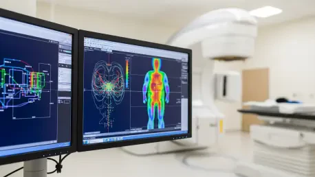
To manage this physical displacement in the past, protocols often required the surgical implantation of small metallic landmarks, known as fiducial markers or “seeds,” into the liver tissue. While these markers are effective for providing a reference point for imaging equipment, the minor surgical procedure required to insert them carries inherent risks such as internal bleeding, organ puncture, or localized infection. The AI technology currently being trialed seeks to replace these physical markers with digital precision, using advanced algorithms to map and monitor the tumor’s movement non-invasively throughout the entire session. By creating a real-time digital twin of the patient’s internal anatomy, the system can account for every inhale and exhale without the patient ever needing to go under the knife for marker placement. This transition from physical seeds to algorithmic tracking marks a major leap in patient safety and comfort during an already stressful period.
Clinical Integration and Regional Research Progress
Validation: Accuracy Through Observational Data Analysis
At this current stage, the trial is functioning in a strictly non-interventional capacity, meaning the artificial intelligence is used to observe and analyze tumor movement during actual sessions without yet taking direct control of the radiation beams. This observational phase is vital for validating the software’s accuracy and ensuring it can reliably predict the tumor’s path as the patient inhales and exhales over several minutes. Lead researchers have noted that the AI’s ability to track these shifts with high precision is the most significant technological milestone achieved since the study began earlier this year. Furthermore, the project demonstrates that regional health centers like Gosford can contribute meaningfully to global medical innovation while providing world-class care close to home. Supported by millions of dollars in government funding, this decentralized research model fosters a safer environment for patients who no longer need to travel to metropolitan hubs for cutting-edge clinical trials.
Implementation: Scaling Automated Treatment for Modern Oncology
Medical professionals looked toward these developments as the bridge between invasive methodologies and a future defined by patient-friendly, automated cancer treatment. As the technology matured throughout the trial, it successfully showed how digital innovation mitigated physical surgical risks while increasing the efficacy of existing hardware. For healthcare providers, the next actionable steps involved integrating these validated algorithms into standard linear accelerators to ensure that every patient benefited from real-time tracking. Policymakers and hospital administrators evaluated the cost-effectiveness of these digital solutions compared to traditional surgical markers and found that the reduction in complications led to better long-term resource allocation. Moving forward, clinical teams prioritized the training of technicians in AI-assisted workflows to ensure that the transition to automated radiotherapy was seamless across all departments. This progress solidified a new era in oncology where precision was no longer a variable.