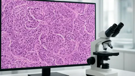

Michigan Medicine’s integration demonstrates that clinical data becomes research-grade when stored in standardized DICOM formats within a vendor-neutral archive. This landmark achievement has set a new standard for medical informatics in the United States, proving that digital pathology is no longer an experimental niche but a foundational element of modern diagnostic workflows. The transition represents a fundamental shift in how diagnostic data is handled, moving from a century-old tradition of physical glass slides to a sophisticated, data-driven digital ecosystem. By leveraging modern standards and scalable storage solutions, Michigan Medicine has demonstrated how pathology can leapfrog the developmental hurdles previously faced by radiology, moving directly into a mature, high-performance digital environment. The institution established a roadmap for how modern healthcare can consolidate diagnostic data to improve clinical precision and patient care.
Breaking Down Data Silos Through Enterprise Imaging
The strategic integration of pathology into an enterprise imaging framework aims to redefine traditional workflows that have historically isolated critical diagnostic information. In the past, pathology images were often confined to a single department, creating data silos that made it difficult for other specialists to access important tissue analysis. Enterprise imaging brings these high-resolution images into the same digital environment as radiology and cardiology, allowing for a unified patient record that is accessible through a single viewer within the Electronic Health Record. By centralizing these assets, healthcare providers can eliminate the fragmentation of patient data that often leads to delays in treatment. When pathology images are part of a broader imaging network, they become more than just static pictures; they transform into dynamic components of a patient’s comprehensive medical history. This holistic approach ensures that any specialist can view the necessary diagnostic evidence.
Historically, the separation of diagnostic departments led to a lack of communication and slower consultation times for complex cases. The current movement toward enterprise imaging seeks to dissolve these silos by creating a shared infrastructure where various imaging modalities coexist. This integration allows for a more fluid exchange of information between departments, fostering a collaborative environment where radiologists and pathologists can share insights more easily. The centralized nature of an enterprise system also simplifies the administrative burden of managing multiple proprietary software systems, as IT departments only need to maintain a single, cohesive platform. As a result, the hospital can focus more resources on patient care rather than troubleshooting disparate legacy systems. This shift not only improves the speed of diagnosis but also enhances the overall quality of care by providing clinicians with a complete picture of a patient’s health status at the point of care.
Technological Foundations: DICOM and VNA Architectures
A successful transition to digital pathology relies heavily on the adoption of Digital Imaging and Communications in Medicine standards for image management. Using DICOM for Whole Slide Imaging ensures interoperability, meaning that scanners, archives, and viewers from different manufacturers can communicate seamlessly without technical barriers. This standardization removes the need for expensive, proprietary translation software and allows hospitals to build a flexible, future-proof infrastructure that can evolve alongside new technological advancements. By adhering to global standards, institutions can ensure that their data remains accessible and usable for decades, regardless of changes in specific hardware vendors. The maturity of these standards in the current year provides a stable foundation for hospitals that were previously hesitant to digitize their labs. This technical uniformity is the bedrock upon which high-performance diagnostic networks are built.
Managing the immense volume of data generated by digital pathology requires a robust storage strategy that balances performance with economic reality. Because tissue scans are significantly larger than traditional radiology files, institutions must utilize Vendor Neutral Archives and tiered storage solutions to remain efficient. This method allows frequently accessed images to remain on high-speed hardware for immediate clinical use, while older cases are moved to cost-effective, long-term storage. This tiered approach ensures that pathologists have the speed they need for active cases while maintaining a complete historical record for the institution. Furthermore, the use of a VNA prevents vendor lock-in, giving the hospital the freedom to switch hardware providers without the risk of losing access to their historical data. By implementing a scalable storage architecture, healthcare systems can manage the data explosion associated with high-resolution imaging while maintaining fiscal responsibility over time.
Artificial Intelligence as a Catalyst for Diagnostic Change
The urgency to digitize pathology is fueled by a growing shortage of specialized pathologists and an increasing demand for complex diagnostic services. Digital workflows allow for greater efficiency, enabling pathologists to share cases instantly for second opinions and manage higher workloads through automated triaging. This speed is essential in modern medicine, where rapid diagnosis can significantly impact the effectiveness of treatment plans for life-threatening conditions. Furthermore, the ability to work remotely or across different hospital campuses allows institutions to better distribute their specialized staff, ensuring that even remote clinics have access to expert analysis. The transition to digital tools also enables the use of advanced annotation and measurement features that are simply not possible with a traditional physical microscope. This digital evolution is not just about convenience; it is about meeting the rising clinical demands of an aging population.
The rise of Artificial Intelligence serves as another powerful catalyst for this digital transformation across the healthcare landscape. AI models require massive amounts of standardized, high-quality data to function effectively, particularly multimodal AI that analyzes radiology and pathology data simultaneously. Without the integrated data structures provided by an enterprise imaging network, these advanced diagnostic tools cannot be fully realized or implemented in a clinical setting. By centralizing diagnostic images, hospitals create a rich dataset that can be used to train and validate AI algorithms that assist in detecting subtle patterns invisible to the human eye. These tools can act as a second set of eyes for pathologists, highlighting areas of concern and reducing the risk of diagnostic errors. As AI continues to advance, the institutions that have already digitized their imaging workflows will be the first to benefit from these sophisticated, life-saving technologies.
Strategic Planning: The Multidisciplinary Blueprint for Success
Integrating pathology into an enterprise network requires a comprehensive, committee-based approach that involves stakeholders from across the institution. Clinical leads, IT professionals, and operations managers must work together to ensure the system meets diagnostic needs while maintaining data integrity and security. Strategic planning must also involve legal and compliance officers to navigate the complexities of patient privacy and data access protocols in a digital environment. One of the most critical lessons from early implementations is that pathology must be an active participant in the design phase from the beginning. If the platform is built without the input of the pathologists who will use it, the resulting workflow may not align with the practical realities of the lab. A multidisciplinary blueprint ensures that the system is not just a technical fix, but a holistic improvement to the operational fabric and the clinical culture of the hospital.
In addition to technical and clinical leadership, successful integration projects often include subcommittees focused on research, faculty enablement, and patient access. This ensures that the benefits of digital pathology extend beyond the diagnostic lab and into the broader academic and patient-facing missions of the organization. For instance, creating a clear pathway for researchers to access de-identified images can accelerate the development of new treatments and clinical trials. Faculty enablement programs can help ease the transition for staff members who are accustomed to traditional methods, providing the training and support necessary to master new digital tools. By addressing these different facets of the hospital’s mission, the strategic planning committee can build a system that is robust, user-friendly, and widely accepted by all staff. This collaborative approach minimizes resistance to change and maximizes the long-term utility of the digital imaging investment.
Practical Implementation: Human Elements and Economic Realities
Technology is only one component of a successful digital transition; the human and organizational elements are equally important for long-term viability. Successful institutions emphasize the need for physician champions who understand both clinical medicine and information technology to act as a bridge between technical teams and medical staff. Executive sponsorship is also vital to secure the necessary funding and overcome the natural inertia that often exists within large, traditional departments. Before implementing a new system, departments must have a clear understanding of their existing and desired workflows to ensure the technology supports the way doctors actually work. A thorough inventory of existing data, including old archives and loose storage devices, is also necessary to understand the full scope of the migration. Without these human-centric strategies, even the most advanced technological systems can fail to gain traction within a clinical environment.
While a standalone pathology system might seem more affordable in the short term, it often leads to significant technical debt that can burden a hospital for years. When an institution eventually decides to integrate a siloed system into a broader network, the migration costs can be prohibitively high, often rivaling the original purchase price. Building on existing enterprise infrastructure results in a lower total cost of ownership over time by avoiding redundant hardware and complex data migrations. By investing in an integrated framework from the start, healthcare systems can create a more sustainable financial model for digital diagnostics. The efficiencies gained through streamlined workflows and the ability to leverage existing IT resources make the enterprise approach a more fiscally responsible choice. As the medical field continues to evolve, the ability to centralize and scale imaging data will remain a key factor in a hospital’s economic and clinical viability.
The Evolving Horizon: Unified Care and Collaborative Outcomes
The successful transition to an integrated digital pathology system demonstrated that the path to modernization was paved with strategic foresight and interdepartmental synergy. Healthcare systems that moved early found that the implementation of unified viewers significantly reduced the time spent on manual slide logistics and preparation. Institutions identified that the most effective first step involved cataloging every existing image source to prevent data loss during the massive migration. The journey proved that while the initial capital expenditure appeared significant, the long-term reduction in technical debt justified the investment for the entire organization. Leaders recognized that fostering a culture of digital literacy among staff members was just as important as the hardware itself for ensuring daily operational success. By the time the integration was complete, clinicians had already begun to leverage the unified database for real-time peer reviews and automated triaging of urgent cases. This shift eventually transformed how diagnostic precision was defined across the entire imaging network.