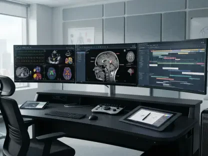

Researchers have recently introduced a fully automated artificial intelligence pipeline designed to eliminate the human bottleneck in quantitative spinal muscle assessment. This development comes at a critical time when clinicians are increasingly recognizing that the structural integrity of the human spine is intrinsically linked to the health of the surrounding paraspinal muscles. These deep-seated muscle groups are the silent guardians of our skeletal system, providing the necessary stabilization for everyday movements and maintaining upright posture against the constant pull of gravity. For years, routine lumbar Magnetic Resonance Imaging has captured detailed snapshots of these tissues, yet the vast majority of this data remains untapped in standard clinical settings. The primary obstacle has always been the sheer logistical burden of manual analysis, which requires highly trained professionals to spend hours tracing muscle boundaries and identifying specific vertebral levels. By transforming this arduous task into an automated workflow, medical technology is finally addressing a long-standing gap in spinal diagnostics, potentially moving muscle health from an afterthought to a primary clinical indicator. This automated approach ensures that high-quality data is available for every patient, regardless of the workload of the radiology department, allowing for more objective and data-driven healthcare decisions in the realm of physical therapy and orthopedic surgery.
Bridging the Gap: End-to-End Automated Pipelines
To prove the system’s efficacy in a realistic setting, the research team utilized data from an external cohort that was entirely separate from the initial training datasets. This methodology is crucial for verifying that the algorithm can perform reliably across the wide variability of real-world clinical environments. Out of 146 eligible cases processed through the new pipeline, the artificial intelligence achieved a staggering 98.6 percent success rate. This level of robustness suggests that the system can handle various patient anatomies and different MRI scanner settings without the common errors that often plague experimental medical software. By successfully navigating these diverse data points, the pipeline demonstrated that it is ready for high-throughput clinical environments where speed and reliability are paramount. The ability to maintain such high accuracy in an external validation set underscores the readiness of this technology for widespread adoption in hospitals that utilize a variety of imaging hardware and patient demographics. This is a significant leap from previous iterations that often required manual adjustments when faced with unfamiliar image quality or unusual anatomical variations, marking a new standard for reliability in automated diagnostic tools.
The technical core of this innovative system relies on the nnU-Net framework, which represents the state-of-the-art in deep learning architectures specifically designed for medical image segmentation. Unlike standard neural networks that require extensive manual tuning for every new dataset, this framework is capable of self-configuring based on the specific characteristics of the images it processes. This adaptability is what allowed the researchers to achieve such high precision across the L3 and L4 vertebral levels. In the context of 2026, such self-optimizing models have become the gold standard for diagnostic tools because they reduce the need for constant developer intervention. The pipeline effectively handles both localization—finding the right spot in the spine—and segmentation—outlining the muscles themselves—in a single, cohesive workflow. This end-to-end automation is designed to fit seamlessly into existing hospital information systems, allowing the results to be generated in the background while the radiologist focuses on identifying other pathologies. This integration is essential for creating a sustainable diagnostic environment where advanced data analysis does not come at the cost of clinical efficiency or increased physician burnout, effectively bridging the gap between cutting-edge research and daily clinical practice.
Validating Precision: Three Performance Domains
The first critical task for the artificial intelligence was the autonomous localization of the L3-L4 vertebral level. This specific anatomical region is widely regarded as the global standard for muscle health assessment because it provides the most representative view of body composition and core stability. In a randomly sampled validation subset, the system achieved a 100 percent hit rate, pinpointing the correct slice with a mean absolute error of just 0.07 slices. Such precision is vital because even a minor deviation of one or two slices can significantly alter the resulting muscle measurements, leading to inaccurate clinical conclusions. By mastering this initial step with absolute consistency, the AI provides a reliable foundation for all subsequent data analysis, ensuring that every measurement is taken from the exact same anatomical plane across different patients. This eliminates one of the most common sources of human error in spinal research, where slight differences in slice selection can lead to high variability in longitudinal studies. The consistency of this automated localization ensures that when a patient returns for a follow-up scan, the comparisons made over time are based on identical anatomical landmarks, providing a true reflection of their progress rather than an artifact of human measurement variability.
Once the correct slice was identified, the AI had to execute the complex task of segmentation by outlining the boundaries of the paraspinal muscles. This performance was measured using the Dice similarity coefficient, a statistical tool where a score of 1.0 represents a perfect overlap with a human expert’s manual tracing. The system achieved an impressive Dice score of 0.934, placing its precision on par with the level of agreement typically found between two experienced radiologists. More importantly, the standard deviation was remarkably tight, indicating that the algorithm maintained its accuracy across patients of all shapes, sizes, and ages. Whether analyzing a young athlete or an elderly patient with significant spinal degeneration, the AI provided consistent and reliable outlines of the muscle tissue. This level of performance is essential for clinical trust, as it proves that the algorithm is not biased toward specific body types or image qualities. The ability to automate this pixel-by-pixel tracing saves hundreds of man-hours in a typical research study and removes the subjective “art” of muscle tracing, replacing it with a rigorous, repeatable mathematical process that can be audited and verified with ease, ultimately leading to more transparent and reliable diagnostic reports for patients and physicians.
Reliability of Measurements: Clinical Implications
The ultimate test of the system’s value lay in its ability to match human calculations for two key biomarkers: Lean Cross-Sectional Area and Fat Fraction. The first metric, LCSA, represents the actual functional muscle tissue available to support the spine, while the Fat Fraction provides an estimate of how much fat has infiltrated the muscle, a condition often referred to as myosteatosis. The Intraclass Correlation Coefficients between the AI’s results and those of human experts ranged from 0.91 to 0.98, falling into the category of “excellent agreement.” These metrics are the bedrock of quantitative imaging, as they are required for any biomarker intended for use in clinical trials or regulatory environments. By providing these numbers with high reliability, the AI allows clinicians to diagnose conditions like sarcopenia—the age-related loss of muscle mass—with much greater objective certainty than a visual inspection would allow. This quantification transforms a qualitative observation of “muscle looks a bit thin” into a precise data point that can be used to guide treatment plans and monitor the effectiveness of rehabilitation programs. Having access to such granular data allows for a more personalized approach to spinal care, where interventions are based on the specific physiological state of the patient’s musculature.
The clinical implications of this automated quantification are far-reaching, especially since the AI is designed to work on standard T2-weighted MRI sequences that are already used for millions of back pain patients annually. This means that a comprehensive muscle health report can be generated without requiring patients to undergo additional scans or incur extra costs. In a typical clinical workflow, a surgeon could use this data to identify at-risk patients who might have poor surgical outcomes due to underlying muscle wasting. For these individuals, a period of pre-operative physical therapy might be recommended to strengthen the back before an invasive procedure is performed, a concept known as prehabilitation. Furthermore, on a broader population level, researchers can now utilize this tool to analyze massive existing databases of spine scans. This allows for a deeper understanding of how muscle health correlates with long-term disability, frailty, and overall longevity. The wealth of “hidden” data already stored in hospital archives can finally be unlocked to provide insights that were previously too labor-intensive to gather. This shift toward data-driven spinal care promises to improve patient outcomes by providing a more holistic view of the factors contributing to back pain and mobility issues.
Future Directions: The Shift in Diagnostic Philosophy
While the results are highly promising, the research team was transparent about the limitations that currently exist within the technology. An exploratory analysis of patients with extreme muscle degeneration revealed that the AI’s fat fraction estimates showed higher levels of uncertainty in these complex cases. When muscle tissue is heavily replaced by fat, the signal intensity within the MRI changes in ways that can challenge even the most advanced deep learning models. This suggests that while the system is exceptionally accurate for the general population, further refinement is needed to ensure peak performance in cases of severe atrophy or rare muscular diseases. Additionally, the researchers noted that the next logical step is to validate the pipeline across a wider variety of medical centers using various MRI scanner brands, such as Siemens, GE, and Philips. Ensuring that the algorithm remains robust across different hardware configurations is a prerequisite for global clinical deployment. By identifying these specific areas for improvement, the study provides a clear roadmap for future development, ensuring that the technology continues to evolve toward total reliability even in the most challenging diagnostic scenarios. This transparency about current limitations reinforces the scientific integrity of the project and sets a clear agenda for the next phase of clinical validation.
The study from the Chongqing Medical University team marked a significant shift in the philosophy of medical artificial intelligence by moving beyond simple decision support toward full task replacement. The researchers demonstrated that an automated pipeline could handle the tedious, high-skill manual labor of muscle assessment faster and more consistently than human experts. This achievement cleared the path for paraspinal muscle health to become a standard vital sign in modern spinal care, much like blood pressure or heart rate. By proving that a “set-it-and-forget-it” system could achieve expert-level results in localization, segmentation, and measurement, the team established a new benchmark for quantitative imaging. Moving forward, the integration of these tools into hospital workstations will allow for the seamless tracking of muscle quality throughout a patient’s life. Healthcare providers should look toward adopting these automated workflows to optimize surgical planning and personalize rehabilitation strategies. As the industry continues to refine these models, the objective assessment of soft tissue health will likely become a cornerstone of preventative spinal medicine. The transition from manual tracing to automated analysis has finally allowed the clinical community to leverage the full potential of existing imaging technology for better patient outcomes and more efficient diagnostic workflows.