
ThinkSono’s technology bridges the expertise gap by allowing nurses and clinicians to record diagnostic-grade recordings without years of specialized sonography training. This breakthrough represents a significant shift in the management of Deep Vein Thrombosis (DVT), a condition that impacts nearly 900,000 Americans each year and causes up to 100,000 deaths. By receiving FDA 510(k) clearance for its guidance software, the medical community has gained a tool that addresses the persistent delays in vascular imaging. Traditional diagnostic methods often require specialized labs that may not be available at all times, especially in rural or underserved areas. This new AI-driven approach allows for the democratization of vascular scans, moving the capability from the specialized lab directly to the patient’s bedside. By empowering a wider range of healthcare professionals to perform these scans, the healthcare system can significantly reduce diagnostic wait times and improve the speed at which life-saving treatments are initiated.
The Technical Framework: AI Guidance and Hardware Integration
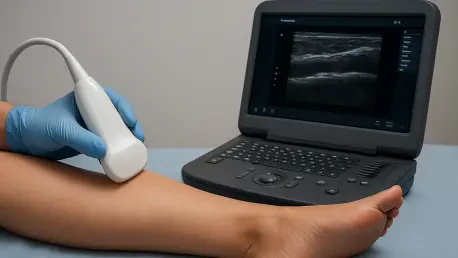
The underlying technology operates by providing real-time, interactive visual cues that guide an operator through the complexities of venous anatomy. When a handheld ultrasound probe is paired with the AI software, the system analyzes the incoming video feed to detect key anatomical landmarks and ensure proper probe compression. It guides the user through the necessary steps to capture high-quality cine-loops of the femoral and popliteal veins, which are the primary sites for identifying deep vein thrombosis. This guidance is particularly valuable for clinicians who may not use ultrasound daily, as it provides a safety net that confirms they are imaging the correct structures. By providing immediate feedback on probe angle and pressure, the AI ensures that the resulting images are of diagnostic quality before the patient leaves the bedside. This technological support effectively turns a standard mobile device into a sophisticated teaching and guidance platform, lowering the barrier to entry for performing critical vascular screenings.
The operational success of this system depends on a human-in-the-loop architecture that balances AI support with clinical oversight. The software does not attempt to automate the final diagnosis; rather, it focuses on the technical challenges of image acquisition. Once the bedside clinician completes the guided scan, the data is instantly transmitted to a secure dashboard for expert review. This workflow ensures that while a nurse can perform the physical exam, a qualified vascular specialist or radiologist remains the final authority on the findings. This separation of tasks effectively removes the specialized labor bottleneck that often stalls DVT diagnosis in the middle of the night or in rural clinics. By allowing the diagnostic workload to be distributed more broadly across the healthcare team, the system allows specialists to focus their time on complex cases and clinical decision-making. This collaborative model has proven essential for maintaining high standards of care while increasing the frequency and speed of diagnostic testing.
Clinical Excellence: Data Validation and Operational Implementation
Clinical validation for this technology was anchored in a substantial multicenter study that evaluated 1,691 patients across 24 distinct clinical sites. The findings were compelling, showing that the software enabled non-experts to achieve a sensitivity of 92.9% and a specificity of 97.1%, which closely aligns with the performance of specialized sonographers. Beyond accuracy, the efficiency of the tool was evident in the median scan time of approximately five and a half minutes. Specialists who reviewed the resulting cine-loops remotely were able to complete their assessments in just over two minutes, demonstrating a streamlined diagnostic lifecycle that is highly suitable for high-volume hospital environments. Most importantly, the AI maintained these high standards of performance across various patient demographics, including differences in Body Mass Index, race, and sex. This consistency is vital for ensuring that the benefits of rapid DVT detection are equitable and reliable for all patients regardless of their physiological characteristics.
The integration of AI-powered guidance into standard hospital workflows provided a blueprint for more accessible vascular care. Facilities that prioritized this transition recognized the immediate benefits of reduced diagnostic wait times and improved resource allocation. To maximize the impact of this technology, healthcare administrators focused on implementing comprehensive training programs for frontline staff, ensuring that 24-hour coverage for DVT screening became a reality. By utilizing existing handheld ultrasound hardware, these organizations avoided significant capital expenditures while expanding their diagnostic reach. The shift toward decentralized imaging protocols demonstrated that high-quality care is achievable through the strategic application of supportive AI tools. Looking forward, the focus remained on standardizing these processes to maintain accuracy across diverse clinical environments. This evolution in practice ensured that patients received timely interventions, ultimately enhancing the overall safety and efficiency of the healthcare delivery system.