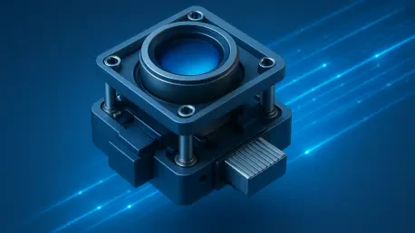

Relying on proprietary cable connectors often creates significant supply chain vulnerabilities for medical device manufacturers who require long-term production stability. In the current landscape of 2026, the shift toward standardized high-performance components has become a necessity rather than a luxury for engineering teams developing the next generation of surgical tools and diagnostic systems. The introduction of the Armor-1335CRO-FPD3 camera module marks a pivotal moment in this transition, offering a sophisticated blend of high-resolution optics and robust connectivity that addresses the most pressing challenges in medical technology. By integrating professional-grade imaging sensors with automotive-style durability, this hardware provides a reliable foundation for instruments that demand extreme precision and consistent uptime. Medical professionals are increasingly looking for visual systems that can bridge the gap between microscopic detail and broad-field observation, and this new module achieves exactly that. As hospitals upgrade their digital infrastructure to support more complex procedures, the hardware must evolve to provide clearer data with less physical bulk. This advancement reflects a broader trend in the industry where the convergence of specialized imaging and standardized hardware architectures is enabling faster development cycles and more resilient healthcare ecosystems worldwide, ultimately improving patient outcomes through better visualization.
Optimizing Visual Fidelity through Advanced Sensor Integration
The core of the Armor-1335CRO-FPD3 module is built around the Onsemi AR1335 sensor, a component that has set a high benchmark for clarity in the current technological environment. This 13-megapixel sensor allows medical devices to capture incredibly detailed still images, which are essential for post-operative documentation and high-precision diagnostic reviews. When surgeons are examining tissue health or identifying minute abnormalities, the pixel density provided by this module ensures that no detail is lost to digital noise or sensor limitations. Beyond static imagery, the module excels in delivering fluid 4K video at thirty frames per second, providing the real-time visual feedback necessary for complex surgical navigation. This dual capability eliminates the need for separate camera systems for documentation and live monitoring, effectively reducing the footprint of the imaging hardware within the surgical suite. By centralizing high-performance video and high-resolution stills in a single unit, manufacturers can design more streamlined devices that do not sacrifice performance for size, a critical factor when every millimeter of space in an operating room is valuable.
The integration of such high-resolution sensors into medical workflows allows for a more nuanced approach to digital pathology and remote consultation. In the current healthcare climate, where specialists often provide guidance from off-site locations, the ability to stream 4K video without significant degradation is a fundamental requirement. The Armor-1335CRO-FPD3 leverages its advanced back-end processing to maintain color accuracy and high dynamic range, ensuring that the visual representation of human anatomy is as close to life as possible. This level of fidelity is particularly important for detecting subtle changes in tissue vascularization or identifying margins during tumor resections. Because the sensor is designed to handle varying light conditions often found in medical settings—ranging from the intense glare of surgical lamps to the dim environments of laparoscopic procedures—it provides a consistent baseline of visual data. This consistency allows for the development of more reliable artificial intelligence algorithms, as the training data and real-time inputs are derived from a high-quality, predictable optical source that minimizes the variables the software must account for during analysis.
Achieving Mechanical Precision with Integrated Stabilization Systems
Maintaining a steady image is one of the most difficult hurdles in medical imaging, especially as surgical procedures become more dynamic and involve multiple moving parts. The Armor-1335CRO-FPD3 addresses this by incorporating Optical Image Stabilization directly into its hardware, which physically compensates for vibrations and movements that would otherwise blur the image. Unlike digital stabilization, which often crops the sensor’s field of view and reduces resolution, the mechanical OIS system maintains the full 13-megapixel integrity of the frame. This is particularly vital when the camera is attached to a robotic arm or a handheld endoscope, where micro-tremors from motors or human hands can significantly degrade the clarity of the visual feed. By neutralizing these disturbances at the source, the module provides a rock-solid image that allows surgeons to maintain their focus on the task at hand rather than being distracted by a shaky or unstable video stream. This physical stability translates directly into higher safety margins during delicate maneuvers where a single blurred frame could lead to a momentary loss of spatial awareness.
In addition to lateral stabilization, the module features a high-speed electronic autofocus system that manages the depth of field with exceptional speed and accuracy. In a medical context, the working distance between the camera and the subject can change rapidly, whether it is a surgeon moving a scope deeper into a cavity or a diagnostic cart being repositioned at the bedside. The autofocus mechanism ensures that the subject remains in sharp focus on the Z-axis, complementing the work done by the OIS on the X and Y axes. This three-dimensional approach to image stability is crucial for maintaining the “immersion” of a surgeon operating through a digital interface. When the focus adjusts seamlessly and the image remains still despite external vibrations, the cognitive load on the medical professional is reduced, allowing them to process visual information more naturally. The combination of hardware-based stabilization and responsive autofocus creates a reliable imaging platform that can adapt to the chaotic and fast-paced nature of modern clinical environments, ensuring that the primary visual data remains crisp and actionable throughout the entirety of a medical procedure.
Strengthening Clinical Hardware with Standardized Interfaces
The adoption of the FAKRA connector and the FPD-Link III interface in the Armor-1335CRO-FPD3 represents a significant departure from the proprietary, fragile connections that have plagued the MedTech industry for years. FAKRA connectors are renowned for their mechanical robustness, featuring a secure locking mechanism that prevents accidental disconnection—a critical failure point during high-stakes surgery. By utilizing a standardized coaxial interface, medical device manufacturers can move away from “single-source” dependencies, allowing them to qualify multiple cable vendors and secure their production lines against localized shortages. This standardization also simplifies the assembly process on the manufacturing floor, as the connectors are color-coded and keyed to prevent incorrect mating. In the fast-paced development cycles of 2026, the ability to rely on industry-standard interconnects means that engineering teams can focus on clinical outcomes rather than troubleshooting bespoke cabling issues, leading to a more efficient path from prototype to commercialized medical device.
The FPD-Link III protocol provides a technical foundation that is perfectly suited for the demanding electromagnetic environment of a modern hospital. Operating rooms are filled with high-frequency equipment, from electrosurgical units to wireless monitoring systems, all of which can interfere with unshielded video signals. FPD-Link III enables the transmission of uncompressed high-definition video, bidirectional control signals, and power over a single, thin coaxial cable that can extend several meters without signal degradation. This “single-cable” solution is a transformative improvement for the design of surgical robots and articulated imaging arms, where minimizing cable bulk is essential for maintaining a wide range of motion. By reducing the number of wires required to operate the camera, designers can create more maneuverable and less intrusive instruments. Furthermore, the protocol’s inherent resistance to electromagnetic interference ensures that the video feed remains clean and lag-free, even when the camera is used in close proximity to other electronic devices, providing a level of reliability that is mandatory for life-critical applications.
Redefining Performance in Robotic Surgery and Endoscopy
In the field of surgical robotics, the camera module serves as the primary sensory link between the patient and the surgeon, who is often positioned at a console several feet away. The Armor-1335CRO-FPD3 provides the low-latency 4K video necessary for this remote interaction to feel instantaneous and natural. Any delay between the surgeon’s hand movement and the corresponding visual update on the screen can lead to “motion sickness” for the operator and, more importantly, a decrease in surgical precision. By optimizing the signal path through the FPD-Link III interface, this module minimizes the time it takes for a photon hitting the sensor to be displayed as a pixel on the monitor. This high-fidelity, real-time feedback loop is what enables surgeons to perform intricate tasks, such as suturing small vessels or dissecting delicate nerve bundles, with the confidence that what they are seeing is an exact, current representation of the surgical field. The 13MP resolution further allows for digital zooming without immediate pixelation, giving the surgeon a closer look at the anatomy when needed.
Endoscopic applications also benefit significantly from the compact and stabilized nature of this imaging platform. Traditional endoscopy often struggled with motion blur as the scope was navigated through narrow, winding internal pathways, but the integration of OIS in the Armor-1335CRO-FPD3 effectively solves this problem. As the endoscope moves, the internal stabilization keeps the view centered and clear, allowing for a more thorough examination of the gastrointestinal tract or respiratory system. The small form factor of the module is another key advantage, as it allows for the development of thinner, less invasive scopes that improve patient comfort and reduce the risk of tissue trauma. By packing 13 megapixels of resolution into such a small footprint, the module enables a level of detail that was previously only available in much larger, more cumbersome camera heads. This advancement allows clinicians to identify smaller lesions and earlier stages of disease, directly contributing to more successful intervention strategies and a higher standard of care for patients undergoing minimally invasive procedures.
Facilitating Mobility through Enhanced Portable Imaging Solutions
The rise of portable diagnostic devices has changed the way healthcare is delivered, bringing high-end imaging to the patient’s bedside, in rural clinics, and even in emergency transport vehicles. The Armor-1335CRO-FPD3 is ideally suited for these mobile applications because its built-in stabilization compensates for the less-than-ideal conditions of field use. When a technician is holding a handheld scanner or a portable microscope, their natural hand tremors can make it difficult to capture a clear image, especially at high magnifications. The OIS system in this module acts as an invisible tripod, smoothing out these movements and ensuring that the resulting image is of diagnostic quality. This democratization of high-performance imaging means that even healthcare workers who are not specialized imaging technicians can produce high-quality results, which can then be shared with specialists via tele-health platforms for immediate assessment. This capability is vital in emergency scenarios where every minute counts and an accurate visual diagnosis can dictate the immediate course of treatment.
For larger, mobile equipment such as medical imaging carts used in hospitals, the standardization provided by this camera module simplifies both the user experience and the maintenance protocols. When a hospital fleet utilizes the same imaging core across various types of carts—whether they are for wound care documentation, intubation assistance, or general nursing rounds—the staff benefits from a consistent interface and predictable performance. This uniformity reduces the amount of training required for nurses and clinicians, as the camera’s behavior, focus speed, and image quality remain the same regardless of which cart they are using. From a facility management perspective, stocking a single type of replacement module or standardized FAKRA cable significantly lowers the overhead costs associated with equipment upkeep. This streamlined approach to hardware ensures that imaging assets are always ready for use, reducing downtime and allowing clinical workflows to remain fluid and uninterrupted, which is essential in high-volume hospital environments where efficiency is directly tied to the quality of patient care.
Ensuring Long-Term Viability in Medical Device Ecosystems
The development of the Armor-1335CRO-FPD3 was driven by a fundamental need for longevity in a sector where product lifecycles often span a decade or more. By selecting automotive-grade components for the sensor and its supporting electronics, the module was built to withstand the rigors of continuous operation and frequent sterilization cycles that are standard in medical environments. This durability helped original equipment manufacturers avoid the frequent redesigns that typically occurred when using consumer-grade parts, which often went obsolete every few years. Manufacturers who adopted this integrated solution found that they could maintain a stable product line from 2026 to 2030 and beyond, without needing to constantly re-validate their optical systems. This long-term availability provided a level of financial and operational security that allowed companies to focus their resources on clinical innovation rather than constant hardware maintenance, ultimately leading to more robust and reliable medical devices in the field.
Looking back at the impact of this technology, the integration of pre-validated camera modules significantly accelerated the regulatory approval process for many new diagnostic tools. Because the stabilization, autofocus, and connectivity had already been rigorously tested as a unified system, engineering teams were able to provide more comprehensive data to regulatory bodies with less effort. This streamlined the path to market, allowing life-saving technologies to reach patients much faster than previous development models allowed. For organizations looking to implement these advancements, the next logical steps involved shifting internal design philosophies away from proprietary silos and toward these standardized, high-performance building blocks. By prioritizing interoperability and component longevity, the MedTech industry successfully reduced the complexity of its supply chains and improved the overall reliability of clinical imaging. This move toward integrated, professional-grade modules proved to be a decisive factor in the successful deployment of advanced robotic and portable medical systems across the global healthcare landscape.