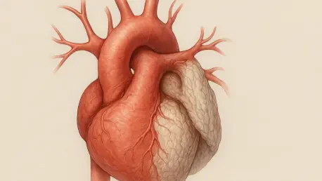

Identifying underlying genetic connective tissue disorders is a vital component of modern aortic care, as these conditions can significantly predispose individuals to arterial wall weakness. This medical reality highlights why aortic aneurysms often operate as shadows within the cardiovascular system, expanding slowly and without warning until a catastrophic event occurs. For many patients, the diagnosis of a localized dilation in the ascending aorta comes as a complete surprise during a routine check-up or an unrelated diagnostic scan, such as a coronary calcium screening. Because these conditions are frequently asymptomatic, they require a meticulous approach to long-term clinical surveillance. Medical professionals emphasize that the absence of pain does not equate to an absence of risk; rather, it underscores the need for a standardized monitoring protocol. By tracking the rate of arterial expansion over several years, clinicians can transform what was once an unpredictable medical emergency into a manageable condition.
The Evolution of Diagnosis and Monitoring
Utilizing Advanced Imaging and Artificial Intelligence
Modern medicine has moved beyond simple observation by integrating high-resolution imaging and artificial intelligence to track arterial changes with unprecedented precision. These advanced technologies allow specialists to measure growth with millimeter-level accuracy, providing a clear, data-driven timeline for when an aneurysm might become dangerous. In 2026, the use of AI algorithms to analyze volumetric data from CT and MRI scans has become the standard of care.
This shift toward precision monitoring ensures that medical teams can wait safely while preparing for the most opportune moment to intervene, rather than relying on guesswork. Furthermore, these diagnostic tools help in identifying subtle changes in the aortic wall that might indicate a higher risk of dissection. By leveraging such sophisticated software, specialists provide patients with a level of security that was previously unattainable in vascular medicine.
Redefining the Surgical Threshold for Patient Safety
While traditional guidelines often suggest waiting until an aneurysm reaches a diameter of 5.5 centimeters, modern specialists are adopting more flexible, patient-centric thresholds to enhance safety. Deciding to operate earlier, perhaps at 5.3 centimeters, can be a strategic move to prevent a rupture or to ensure that the patient’s native anatomy is still healthy enough for advanced repair techniques. This proactive timing is essential for moving from passive monitoring to active surgery.
Factors such as the rate of growth, the patient’s overall health, and specific genetic markers now play a larger role in determining the surgical window than a single measurement. Experts have found that a planned, elective procedure at a specialized center can reduce mortality to less than one percent, which is a stark contrast to the extreme risks associated with emergency operations. Consequently, the focus has shifted to individual risk profiles to ensure the highest survival rates.
Integrated Care and Surgical Innovation
The Benefits of a Multidisciplinary Medical Team
The most effective way to manage complex aortic issues is through a comprehensive care model that brings together various specialists under one roof to coordinate treatment. By combining the expertise of cardiothoracic surgeons, cardiovascular imaging specialists, and geneticists, a hospital can create a unified treatment plan tailored to the unique physiological needs of the individual. This collaborative approach ensures that every aspect of the patient’s health is addressed with specialized precision.
In this environment, the transition from surveillance to surgery is seamless, as the team already possesses a holistic understanding of the patient’s history. This model also facilitates better communication between doctors and families, providing clarity during high-stress decision-making periods. Such integration has proven vital for identifying non-obvious complications early, significantly improving the safety and success of complex interventions across diverse patient populations.
Advancing Patient Outcomes with Valve-Sparing Techniques
A major breakthrough in aortic surgery was the ability to perform valve-sparing procedures, where the patient’s own heart valve was preserved and reimplanted into a synthetic graft. This technique proved particularly beneficial for younger patients, as it maintained natural cardiac function and eliminated the lifelong need for blood-thinning medications. By utilizing these innovative surgical methods within a multidisciplinary framework, medical centers restored the health of many and offered a normal life.
Looking ahead, the focus shifted toward universal screening for those with a family history of arterial disease. Proactive steps included the adoption of genetic testing as a routine part of the diagnostic workup to identify vulnerable individuals before an aneurysm even formed. These strategies effectively transformed the management of “silent” conditions from a reactive struggle into a proactive victory. Specialists recommended that any individual with risk factors sought early imaging for security.