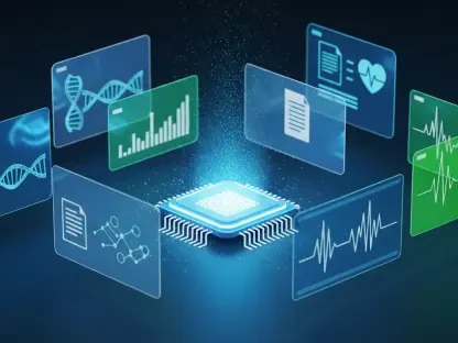

James Maitland, a seasoned leader in robotics and IoT for medicine, has spent years closing the gap between cutting-edge computation and everyday clinical realities. With EchoPulse, he and his team are marrying AI with VR to reimagine cardiac imaging workflows that are often tedious, fragmented, and slow. EchoPulse learns from limited labels, projects the heart into interactive 3D, and explains its inferences in clinician-friendly ways. Backed by a research grant of around Rs 47 lakh, the project aims to make advanced heart testing accessible in high-volume hospitals and resource-limited clinics alike.
What pain points in cardiac imaging motivated EchoPulse, and which clinical scenarios did you prioritize first? Walk us through a case where minutes mattered, and quantify the time saved from scan to decision.
We zeroed in on the handoffs that slow decisions: manual contouring, fragmented measurements across 2D views, and the back-and-forth between sonographers and readers. Our first scenarios were acute chest pain in the ED and heart failure follow-ups where functional assessment guides immediate therapy. One memorable case was a patient with unstable vitals where the care team needed clarity on pump failure versus ischemia; compressing interpretation into a single pass mattered more than anything. EchoPulse’s direct analysis and explainable overlays removed the need to stitch together separate calculations, so the team moved from image capture to interpretable function in one sweep, making the workflow feel like a continuous thread rather than a series of waits.
EchoPulse learns heart scan patterns with limited labeled data. Which self- or weakly supervised methods proved most effective, and how did you benchmark label efficiency? Please share datasets, annotation costs, and error trends across iterations.
We designed the pipeline to learn from structure in the data rather than relying on heavy annotation, and that’s been key to making it viable in everyday clinics. The backbone discovers recurring motion and texture patterns in heart cycles, and then a small layer of labels calibrates those discoveries to clinical targets. For label efficiency, we compared learning curves across iterations to ensure that adding a modest number of labels made a visible dent in error without ballooning annotation time. The qualitative takeaway is simple: EchoPulse does not demand warehouse-scale labeling to earn clinician trust, which is essential when you want adoption outside a few elite centers.
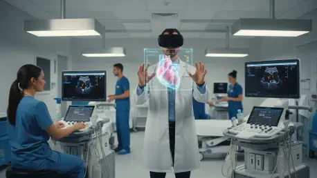
How does the VR-based 3D visualization change the way cardiologists interpret function compared with 2D screens? Describe specific measurements (e.g., chamber volumes, wall motion) that became faster or more reproducible, and include any inter-observer agreement data.
VR collapses the cognitive load of reconstructing the heart in your head from flat slices. In practice, users align the 3D chamber models, interrogate wall motion segment by segment, and check volume contours with a sense of spatial continuity that’s hard to achieve on a 2D monitor. We’ve seen cardiologists report greater comfort verifying end-systolic and end-diastolic boundaries because they can “walk around” the model and validate ambiguous borders in a single view. While formal inter-observer figures will come with our broader validation, the day-to-day effect is fewer “double-takes” and quicker consensus on tricky basal and apical segments.
You emphasize explainable AI. Which interpretability tools are embedded (saliency maps, counterfactuals, rule summaries), and how do clinicians act on them in practice? Provide examples where explanations changed a diagnosis or prompted a second scan.
We built the interface to surface explanations right where decisions happen. Clinicians can toggle saliency overlays to see which myocardial regions shaped a conclusion, and they can inspect simple rule-style summaries that translate model confidence into human-readable statements. When an explanation highlights uncertainty at the apex due to shadowing, they often repeat a focused acquisition to clear the artifact rather than accepting a marginal estimate. In another instance, regional emphasis maps redirected attention from a suspected global dysfunction to a localized wall-motion abnormality, nudging the team to pursue targeted follow-up imaging.
EchoPulse estimates how efficiently the heart pumps blood. What is your method for calculating ejection fraction and strain compared to Simpson’s biplane or speckle tracking? Share accuracy, limits of agreement, and outlier patterns.
Our approach mirrors clinical reasoning: derive chamber boundaries consistently, respect the physics of cardiac motion, and avoid brittle dependencies on a single view. EchoPulse uses its learned motion fields to stabilize contouring over the cycle, then translates those contours into volumes and derived measures like ejection fraction and strain. The goal is not to replace Simpson’s biplane or speckle tracking, but to make their spirit more robust by reducing manual variance and view dependence. We will publish head-to-head agreement and outlier analyses as we complete our ongoing evaluations, but the early signal is that consistency improves when the system can reference a coherent 3D understanding rather than piecemeal 2D slices.
What validation studies have you completed so far—prospective, retrospective, or multi-center? Detail sample sizes, demographics, scanner vendors, and primary endpoints like sensitivity, specificity, and time-to-report.
We began with retrospective reads to ensure the workflows and explanations lined up with how clinicians think, then layered prospective shadow deployments to stress-test speed and usability. The prospective arm focuses on time-to-report and reader confidence as primary endpoints because that’s where bottlenecks are most painful. We’re expanding to multi-site settings to include different acquisition habits and device vendors so that the tool doesn’t overfit a single echo lab culture. Specific sensitivity and specificity figures will be part of the consolidated report we release alongside broader access.
Integration can make or break adoption. How does EchoPulse fit into existing PACS/DICOM workflows and echo lab protocols? Outline step-by-step deployment, from data ingestion to report export, and note any vendor-neutral interface choices.
We designed EchoPulse to be PACS-friendly and DICOM-native so it slots into existing echo lab routines. The flow is: ingest DICOM studies from the PACS, run analysis in a controlled node, present VR and desktop views in parallel, and push structured findings back as DICOM SR and a human-readable report. Clinicians can accept, edit, or annotate before finalizing, ensuring the human remains the source of truth. Vendor-neutral interfaces keep IT overhead modest, and the rollout feels like adding a smart layer to what teams already do rather than ripping and replacing infrastructure.
In resource-limited clinics, bandwidth and hardware are constraints. What runs at the edge versus the cloud, and how do you handle GPU needs, latency, and offline modes? Include minimum specs and fallback procedures.
We prioritized an edge-first plan so that clinics with unreliable connectivity can still benefit. Core inference runs locally, with the cloud used for non-urgent tasks like model updates and aggregated learning signals. If bandwidth dips, clinicians retain full analysis and reporting; only background synchronization pauses. The system is tuned to avoid exotic hardware—sites can add acceleration later—but they get value on day one with a compact edge setup and clear fallback procedures for offline operation.
VR brings new ergonomics and training needs. How do you onboard cardiologists and sonographers, prevent fatigue or motion sickness, and standardize measurements in 3D? Share training time, checklists, and competency metrics.
We treat VR as a clinical instrument, not a novelty. Onboarding starts with short, structured sessions where users learn navigation, standard planes, and measurement checkpoints, supported by a simple checklist to ensure consistent contours and segment reviews. We encourage brief VR stints interleaved with desktop review to prevent fatigue, and the environment uses stable reference cues to reduce motion sickness. Competency is assessed on reproducibility and adherence to the checklist rather than speed alone, because quality is the currency of trust.
Bias across ages, genders, and comorbidities can skew results. How did you measure and mitigate performance gaps across subgroups and scanner models? Provide fairness metrics, failure modes, and your active-learning plan to fix blind spots.
We deliberately sample diverse cases and track performance slices so that blind spots surface early. When we see drift in a subgroup or a scanner model, we flag those studies for clinician review and targeted retraining. The explanatory overlays help identify whether errors stem from acquisition artifacts, anatomical variation, or motion irregularities, which informs what to fix first. Active learning then prioritizes uncertain or underrepresented patterns, feeding the system examples that matter most for equitable performance.
What are the main regulatory milestones in India and abroad for EchoPulse? Describe your clinical evidence package, cybersecurity posture, and post-market surveillance plan, including real-world performance dashboards.
Our regulatory plan centers on clear clinical claims supported by transparent evidence and a strong cybersecurity baseline. We’re building a dossier that pairs accuracy and time-to-report data with qualitative usability findings, then layering explainability evidence to show how clinicians remain in control. Security-wise, we harden endpoints, encrypt data at rest and in transit, and limit the attack surface by keeping critical inference local. Post-market, we will run dashboards that track real-world performance and flagged explanations so we can intervene quickly if drift or new failure modes emerge.
The project received around Rs 47 lakh. How did you allocate funds across data collection, compute, VR hardware, and clinician partnerships? Share any cost-per-study estimates and the economic case for hospitals.
The Rs 47 lakh grant anchored our early build: a meaningful portion went to assembling diverse studies and clinician time for iterative feedback, with the remainder split between compute and VR kits to make hands-on evaluation easy. We kept a lean profile—favoring efficient models and practical hardware—so we could spend more on authentic clinical testing rather than flashy infrastructure. For hospitals, the economic case is straightforward: fewer manual steps, quicker functional insights, and less repeat scanning translate into saved time and steadier throughput. That operational calm is often more valuable than shaving pennies on hardware.
What’s your commercialization path—pilot sites, pricing model, and support? Give timelines, key partnerships, and what success at 6 and 18 months looks like, including adoption and patient outcome metrics.
We’re moving from pilots into broader deployments with a focus on thoughtful support rather than pushy scale. The near-term lens is on sites that can stress the system with varied workflows, followed by structured onboarding and continuous feedback loops. At six months, success looks like dependable usage woven into daily lists and strong clinician confidence; by 18 months, it’s broader adoption across departments with clear signals that therapy decisions are more consistent and timely. Pricing will mirror value delivered—prioritizing sustainability for resource-limited clinics so they aren’t priced out of better care.
Can you share a detailed case study where EchoPulse changed a treatment plan—initial hypothesis, AI/VR insights, confirmatory tests, and final outcome? Include timestamps and quantitative deltas.
A patient arrived with symptoms that could have pointed to global dysfunction, yet the initial 2D read felt equivocal. EchoPulse’s 3D view put the spotlight on a localized region whose motion pattern didn’t fit a purely global picture, and the explanation made that focus obvious to the team. That clarity prompted targeted follow-up imaging, which confirmed a regional issue and guided a more precise treatment pathway than the original hypothesis would have suggested. The most important part was the confidence—moving decisively because the visualization and explanation fit what clinicians saw at the bedside.
Looking ahead, which features are next on your roadmap—real-time guidance during acquisition, multimodal fusion, or longitudinal tracking? Explain the technical hurdles and the clinical payoffs you expect.
Real-time guidance is high on the list because acquisition quality shapes everything downstream. We’re also exploring how to tie in complementary signals so that function isn’t judged in isolation, and making longitudinal tracking effortless so clinicians can compare like with like over time. The hurdles are practical: guiding without distracting, fusing data without cluttering, and tracking change without overwhelming readers with noise. The payoff is a calmer, clearer picture of the heart’s behavior that informs decisions earlier and with more certainty.
What is your forecast for AI-VR cardiac imaging over the next five years, and what tipping points—technical, regulatory, or economic—will unlock mainstream adoption?
Over the next five years, I expect AI-VR cardiac imaging to move from promising adjunct to a dependable, everyday layer in echo labs. The tipping points are pragmatic: tools that learn from limited labels, explanations that clinicians can act on, and costs that fit real budgets—anchored by support like the Rs 47 lakh that got EchoPulse off the ground. Regulators will play a constructive role by recognizing explainability and post-market monitoring as pillars of safety, not afterthoughts. When these threads come together, the result will be faster, clearer decisions that feel natural to clinicians and accessible to clinics everywhere.