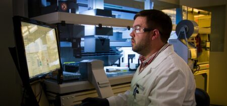


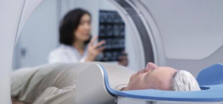


Interventional radiologists at Stanford University Medical Center are using visualization software from EchoPixel that turns 2D CT scans into 3D images so they can virtually view patents’ unique arterial anatomy to help them prepare for endovascular repair of splenic artery aneurysms.
According to Zlatko Devcic, MD, a fellow of interventional radiology at Stanford University School of Medicine, splenic artery aneurysms—a rare and life-threatening clinical disorder—have complex anatomy that require meticulous pre-procedure planning.